
LowKV STEM
Contact: Dr. Erich Müller
In electron microscopy there is a strong demand for information regarding the thickness and composition of the samples. Scanning transmission electron microscopy (STEM) at low energies (up to max. 30keV) provides an approach to these properties. State of the art scanning electron microscopes (SEM) equipped with a standard STEM detector are used both for imaging and analyzing the transmitted electrons. By comparison with simulated or calculated fractions of transmitted electrons, the thickness and composition of the sample can be determined with high lateral resolution. For additional accuracy samples with defined thickness or wedge angle are prepared by means of focused ion beam (FIB) techniques. The use of SEM typical low energies allows to examine even specimens, which are extremely sensitive towards knock-on damage.

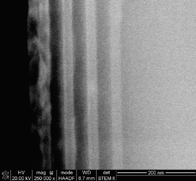
The samples on which our research is focused at present consist of InGaAs layers in a GaAs matrix. The InGaAs layers have a thickness of 20 nm and are separated by GaAs barrier layers of 35 nm thickness. The In-concentration in the layers is varied from 10 % - 40 %, as shown in the schematic cross-section view on the left-hand side.
A STEM image taken in the HAADF (high-angle annular dark-field) mode of a conventionally prepared cross-section TEM sample is shown. The layers can be well distinguished due to the different In-concentrations which lead to different intensities in the image. The forth layer with 40% In shows additional contrast resulting from defects. The intensity of the GaAs barrier and buffer layers increases due to the increasing TEM sample thickness.
Selected conference poster presentations:
Sample thickness determination by low-energy scanning transmission electron microscopy (pdf)
Quantitative Transmission Measurements in a Scanning Electron Microscope (pdf)
Selected publications:
[1] T. Volkenandt, E. Müller, D. Gerthsen, D. Hu, D. Schaadt,
Quantification of the In concentration of InGaAs quantum wells by transmission measurements
in a scanning electron microscope, in W. Grogger, F. Hofer, P. Pölt (Eds.),
Proc. MC2009 Microscopy Conference, Graz, Austria (2009),
Vol. 3: Materials Science, p. 295, Verlag der TU Graz 2009
[2] T. Volkenandt, E. Müller, D. Hu, D. Schaadt, D. Gerthsen,
Quantification of Sample Thickness and In-Concentration of InGaAs Quantum Wells by Transmission Measurements
in a Scanning Electron Microscope,
Microscopy and Microanalysis 16, 604-613 (2010)
[3] M. Pfaff, E. Müller, M. Klein, A. Colsmann, U. Lemmer, V. Krzyzanek, R. Reichelt, D. Gerthsen,
Low-energy electron scattering in carbon-based materials analyzed by scanning transmission electron microscopy
and its application to sample thickness determination,
J. Microscopy 243, 31 (2011)
[4] M. Pfaff, E. Müller, M. F. G. Klein, A. Colsmann, U. Lemmer, V. Krzyzanek, R. Reichelt and D. Gerthsen.
Low-energy electron scattering in carbon-based materials analyzed by scanning transmission electron microscopy
and its application to sample thickness determination.
Journal of Microscopy 243, 31-39 (2011)
[5] M.F.G. Klein, M. Pfaff, E. Müller, J. Czolk, M. Reinhard, S. Valouch, A. Colsmann, U. Lemmer, D. Gerthsen,
Poly(3-hexylselenophene) solar cells: Correlating the optoelectronic device performance and nanomorphology
imaged by low-energy scanning transmission electron microscopy,
J. Polymer Science: Polymer Physics 50, 198 (2012)
[6] M. Pfaff, M.F.G Klein, E. Müller, P. Müller, A. Colsmann, U. Lemmer, D. Gerthsen,
Nanomorphology of P3HT:PCBM-based absorber layers of organic solar cells after different processing
conditions analyzed by low-energy scanning transmission electron microscopy,
Microsc. Microanal. 18, 1380 (2012)
[7] H. Gehrke, A. Frühmesser, C. Marquardt, J. Pelka, M. Esselen, L. Hecht, H. Blank, H.P. Schuchmann, D. Gerthsen,
S. Diabaté, C. Weiss, D. Marko,
In vitro toxicity of amorphous silica nanoparticles in human colon and lung carcinoma cells,
Nanotoxicology 7, 274 (2013)
[8] H. Blank, R. Schneider, D. Gerthsen, H. Gehrke, K. Jarolim, D. Marko,
Application of low-energy scanning transmission electron microscopy for the study of Pt nanoparticle
uptake in human carcinoma cells,
Nanotoxicology 8, 433 (2014)
[9] T. Volkenandt, E. Müller, D. Gerthsen,
Sample thickness determination by scanning transmission electron microscopy at low electron energies,
Microsc. Microanal